Histology and Cytology Unmasked: Decoding The Secrets Of Cellular Life
Histology and Cytology plays a crucial role in various scientific disciplines, including cell biology, genetics, pathology, and reproductive biology. By investigating cells, cytologists can gain insights into the mechanisms underlying diseases, such as cancer, and develop novel therapies. They can also analyze cellular samples for diagnostic purposes, aiding in the detection and classification of diseases. Cytologists employ a range of techniques to study cells. They use staining methods to visualize cellular components, such as the nucleus, cytoplasm, and organelles. Specialized techniques like immunocytochemistry allow the identification of specific proteins within cells.
Flow cytometry enables the analysis of thousands of cells simultaneously, providing valuable information about cell populations and their characteristics. Histology and Cytology are interconnected disciplines that complement each other. While histology focuses on tissues, it relies on cytology to examine individual cells within those tissues. Cytology, in turn, benefits from histological knowledge to understand the cellular organization and context. Histological analysis provides cytologists with valuable information about the tissue environment, enabling them to interpret cellular features more accurately.
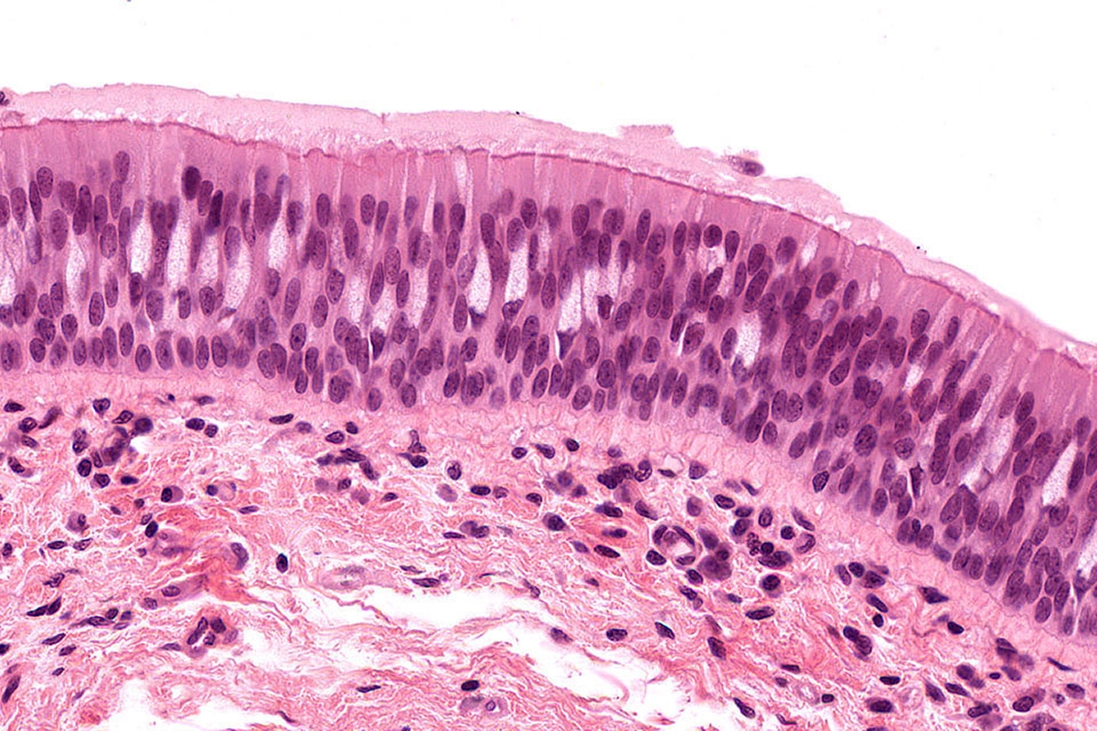
Histological studies rely on the use of specialized stains and dyes that selectively target different components of tissues, aiding in their identification and differentiation. Hematoxylin and eosin (H&E) staining, for example, is widely used in histology to visualize nuclei (hematoxylin stain) and cytoplasm (eosin stain) in tissue samples. This staining technique facilitates the identification of different cell types and the overall architecture of tissues.