External Fixators: An Innovative Solution for Bone Fixation
What are External Fixators?
They are medical devices used in orthopedic surgery to stabilize bones and joints temporarily. They provide stabilization on the outside of the body and can be adjusted as the body heals. They help hold together fractures or broken bones so they can heal properly without needing to undergo internal fixation surgery.
How do They Work?
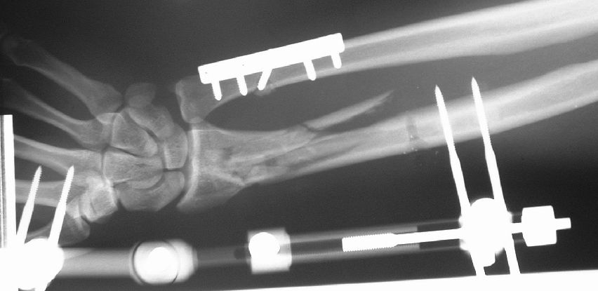
They work by connecting bone fragments External Fixators using articulated joints, rods, wires and pins/screws. Specially designed pins or screws are drilled into the bones above and below the fracture site. The pins are then connected on the outside of the body to metal rods, clamps or rings. This forms a rigid construct around the bone fragments to help keep them aligned during healing. The pins protrude from the skin but are adjusted to avoid putting pressure on nearby nerves, blood vessels or other tissues. They allow flexible stabilization that surgeons can adjust noninvasively as the fracture consolidates.
Types
There are different types of external fixators based on their design, materials and intended clinical use:
Uniplanar or monolateral fixators - Consists of one bar parallel to the bone with pins inserted in one plane on either side of the fracture. Used for simple fractures with small bone segments.
Circular fixators - Have either full or partial rings joined by struts or rods where pins are mounted along with distraction/compression devices. Allows multifocal corrections in different planes with maximum stability and flexibility. Commonly used for limb lengthening and complex fractures.
Hybrid external fixators - Combine features of circular and uniplanar fixators by providing a stable construct in multiple planes using bars in different orientations joined by connectors. More rigid than uniplanar fixators.
Ilizarov fixator - A type of circular external fixator pioneered by Russian orthopedic surgeon Gavriil Ilizarov to treat complex fractures, nonunions and bone defects. Features a series of individually adjustable, carbon fiber rings interconnected by thin wires under tension. Very stable and versatile to slowly redirect bones and lengthen or shorten limbs.
Half-pins vs wires - Different types of fixation attachments used which offer variable hold and stiffness. Pins provide greater stability while wires minimize soft tissue trauma and allow for angled insertions but are less stable. The choice depends on fracture type and bone quality.
Advantages
There are several key advantages of using it compared to traditional internal fixation methods:
- Minimally invasive - They minimize soft tissue dissection required as pins/wires provide least tissue damage and blood loss compared to plates and screws. This reduces surgical trauma and healing time.
- Adjustable construct - As bones heal and swelling subsides, the fixator frame can be noninvasively adjusted under imaging guidance if needed to maintain reduction. This fine tunes the stabilization as healing progresses.
- Stable fixation - Properly applied they are very rigid and provide absolute stability to securely hold even severely displaced fracture segments in correct alignment. Internal fixation has more micromotions at fracture site.
- Preserve soft tissues - Less surgical soft tissue stripping preserves blood supply to bone fragments and tissues. This minimizes complications like infection, nonunion and muscle/nerve damage compared to extensive internal surgeries.
- Frame visibility - The external fixator device sits outside skin allowing direct visualization of pins as infection risks. Any loosening, discharge or pain around pins is immediately noticeable.
- Fracture access - They enable access to complex and comminuted fractures that are difficult to address internally due to hardware constraints, poor bone quality or extensive soft tissue damage.
- Temporary solution - Serves as definitive or temporary stabilization that can later be exchanged for internal implants once soft tissues heal and swelling subsides, allowing less invasive plating. Its removal avoids risks from retained hardware.
Potential Limitations and Complications
While external fixation has many advantages, certain limitations and complications can also arise:
- Pin track infections - Entry points where pins penetrate skin have a risk of developing pin track infections which can spread to bone. Careful sterility and regular cleaning is needed.
- Loosening pins - As fixators rely completely on integrated pins for stability, loss of purchase due to pins backing out over time reduces construct rigidity. Stable fixation requires optimal pin placement.
- Pain and discomfort - Presence of pins protruding through skin can cause irritation, pain with movement and limit ranges of motion during early healing stages before replacement with internal hardware.
- Nonunion risks - Comminuted or severe fractures may not achieve rigid fixation with external devices alone increasing likelihood of delayed or non-unions developing without supplemental bone grafting.
- Limb length issues - Without precise techniques, fixators can introduce unintended limb shortening or lengthening errors due to micromotions at fracture site impacting overall length.
- Pin site infections difficult to treat - Once pins become infected deep within bone, it necessitates more invasive irrigation, debridement and retention of it to eradicate infections. Total eradication delays overall treatment.
external fixators are an innovative solution for temporary fracture stabilization and bone alignment in situations necessitating minimal soft tissue stripping or complex multiplanar reduction. However, their use requires additional precautions due to risks from pin tract infections and reliance on optimal pin fixation.
Get More Insights on- External Fixators
About Author:
Ravina Pandya, Content Writer, has a strong foothold in the market research industry. She specializes in writing well-researched articles from different industries, including food and beverages, information and technology, healthcare, chemical and materials, etc. (https://www.linkedin.com/in/ravina-pandya-1a3984191)