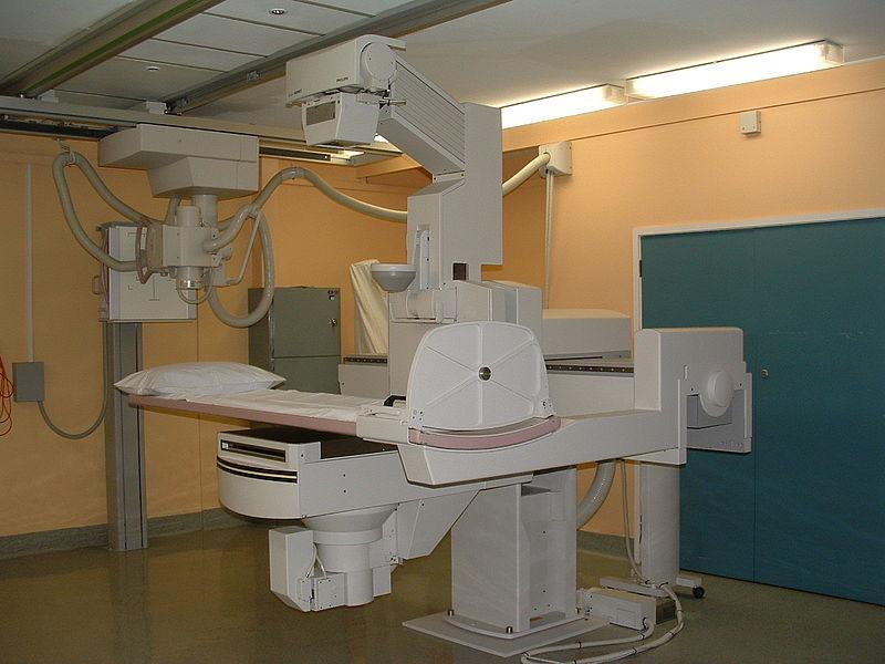
Digital Fluoroscopy Systems: Enhancing Interventional Procedures through Advanced Imaging Technologies
Digital fluoroscopy systems provide real-time moving x-ray images that are displayed on a monitor. Unlike traditional fluoroscopy which uses x-ray film, digital fluoroscopy uses a digital imaging chain and flat panel detectors to capture and display images. This allows interventional physicians to guide medical devices and perform procedures while continuously monitoring progress on high resolution monitors in real time. Digital technologies have enhanced capabilities for interventional imaging compared to traditional fluoroscopy.
Advantages of Flat Panel Detectors
One of the main components enabling digital fluoroscopy is the flat panel detector. Traditional fluoroscopy uses image intensifiers which have limitations in image quality compared to modern flat panel detectors. Flat panel detectors capture x-ray images using an array of tiny pixels similar to a digital camera sensor. Each pixel records x-ray information and sends it to the imaging computer for processing and display. Flat panel detectors provide higher resolution images than image intensifiers for better visualization of anatomy and devices. They also have faster image capture speeds enabling live motion imaging without blurring. This helps physicians accurately guide devices in real time. Flat panels also allow larger viewing areas onmonitors compared to the limited sizes of image intensifier displays.
Image Processing and Display Capabilities
Digital Fluoroscopy Systems imaging chains in modern systems offer advanced image processing features. Images can be digitally filtered and processed to optimize contrast, brightness and other display parameters depending on the clinical need. Various digital processing modes like roadmapping, magnification and digital subtraction enhance visualization. Roadmapping overlays a live fluoroscopy image over previously captured images to help track instrument navigation. Digital subtraction eliminates static anatomy to better visualize moving devices. Images can also be captured, stored, retrieved and compared side by side on high resolution monitors. Physicians have access to advanced visualization capabilities for improved guidance during complex interventional procedures.
Reduced Patient and Staff Radiation Exposure
One significant benefit of digital fluoroscopy compared to traditional systems is lower radiation exposure for patients and staff. Image processing features like automatic dose control and last image hold aid in minimizing unnecessary radiation. Automatic dose control systems constantly monitor dose rates and modulate power output based on real time measurements to use only as much radiation as needed. Last image hold allows physicians to pause live imaging and refer to the most recent capture, reducing cumulative dose over long procedures. Flat panel detectors require lower doses compared to image intensifiers for equivalent image quality levels. combined with dose control technologies, digital fluoroscopy achieves substantial reductions in radiation exposure. This enhances safety for patients especially those undergoing multiple interventional exams. Lower radiation also benefits medical personnel performing procedures on a daily basis.
Expanding Applications of Digital Fluoroscopy
As image quality progressively improves with digital technologies, new interventional applications are emerging. Traditional uses include procedures like angiography, balloon angioplasty and stent placements. More recently, complex endovascular therapies for conditions like aortic aneurysm, carotid artery disease and stroke intervention utilize digital fluoroscopy guidance. Minimal access spine surgery with navigated instruments also leverages real time imaging. Newer applications continue advancing to less invasive treatments for structures like peripheral arteries, abdominal blood vessels, cardiac structures and pediatric interventions. Rapid acquisition interventional CT also integrates real time imaging for novel applications combining morphological and functional imaging guidance. The expanding scope of minimally invasive therapies relies on the enhanced capabilities offered by modern digital fluoroscopy systems.
Integration with 3D Imaging Modalities
Digital fluoroscopy is increasingly being combined with 3D vascular and anatomical imaging modalities to offer comprehensive intra-procedural guidance. Systems allow co-registration of pre-procedure CT/MRI information with real time 2D images for improved navigation and device localization in 3D space. Physicians have access to both 2D live images as well as supplemental offline 3D roadmaps derived from prior scans. This aids complex treatment planning directly in the angio suite or OR. Certain advanced systems provide true 3D fluoroscopy by rotating the x-ray source, enabling the visualization and subtraction of 3D vessels from live cases. Hybrid vascular/interventional suites also integrate digital fluoroscopy with complementary imaging such as CT or ultrasound for multi-modality guidance. Combined with 3D modalities, current digital fluoroscopy platforms offer a more robust and versatile imaging guidance solution.
Implementation Considerations
While digital fluoroscopy enhances low level capabilities over conventional systems, certain implementation factors need attention for optimal clinical benefit. Upgrading requires financial investment along with infrastructure considerations related to equipment footprint and installation requirements. Integration into existing imaging departments and interventional suites demands diligent planning. Operator training on new system functionality and capabilities is crucial, especially advanced 3D/2D fusion tools. Workflow redesign may be needed to capitalize on new guidance options like simultaneous angiographicruns or volumetric roadmaps. Appropriate choice of systems and options tailored to individual institution needs and case mix yields the most advantageous outcomes. Post-installation performance monitoring and ongoing staff education expands utilization over time. With diligent implementation, digital fluoroscopy significantly improves procedural consistency, quality and safety.
Digital fluoroscopy has transformed interventional imaging through state-of-the-art flat panel detectors, advanced processing, expanded display and reduced radiation exposure. Enhanced 2D guidance along with integration of complementary 3D datasets provides comprehensive real time visualization solutions. Continued technology innovations will further optimize image quality, dose efficiency, 3D capabilities and new applications. When implemented judiciously through infrastructure support and operator training, digital fluoroscopy platforms maximize clinical and economic benefits for both patients and institutions. They are revolutionizing minimally invasive interventions by facilitating precise, real time therapy guidance.
Get more insights on Digital Fluoroscopy Systems
About Author:
Money Singh is a seasoned content writer with over four years of experience in the market research sector. Her expertise spans various industries, including food and beverages, biotechnology, chemical and materials, defense and aerospace, consumer goods, etc. (https://www.linkedin.com/in/money-singh-590844163)
