The Advancements in Medical Imaging Phosphor Screen Scanner Revolutionizing X-Ray Technology
Medical imaging technology has come a long way in aiding physicians to accurately diagnose diseases. One such innovation that is set to transform digital radiography is the phosphor screen scanner. Traditional x-ray films use phosphor screens to capture the x-ray image which are then developed into prints. Phosphor screen scanners digitize this process, offering significant benefits to both patients and radiologists.
What are Phosphor Screens?
Phosphor screens are materials coated on thin plastic or glass substrates that emit visible light when exposed to x-rays. The most common phosphor used is calcium tungstate doped with other elements. When x-rays hit the phosphor particles, they create electron-hole pairs which recombine, releasing photons of visible light. The intensity of light emitted is directly proportional to the x-ray exposure. This light image embedded in the phosphor screen is what forms the latent x-ray image.
Traditional Methods and Drawbacks
Conventionally, the phosphor screen containing the x-ray image would then be developed into a film using chemical solutions. This photostimulable phosphor (PSP) plate had to be taken to a darkroom for processing. It involved numerous wet chemistry steps and produced a physical film. Alternatively, cassettes containing screens could be loaded into dedicated film processors for automatic development. However, both methods produced analogue images requiring further digitization for storage and transmission. They were labor intensive, produced chemical waste and had limited image manipulation capabilities.
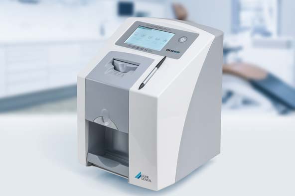
How Phosphor Screen Scanners Work
Phosphor screen scanners digitize the light image embedded in the phosphor screen non-destructively, without the need for wet processing. The screen is loaded into the scanner where a laser beam scans across its entire surface. The laser causes the stored light in the phosphor particles to be emitted. A photodetector senses this stimulated light emission and converts it into digital signals. Sophisticated algorithms then reconstruct this data into a high resolution 16-bit digital radiographic image file.
Key Benefits of Phosphor Screen Scanners
- Filmless Workflow: Scanned images can be viewed, stored and transmitted digitally instead of physical films. This streamlines the workflow.
- Instant Imaging: Images are available instantaneously at the point-of-care without any delay for chemical processing.
- Enhanced Diagnosis: 16-bit grayscale images offer over 65,000 shades of gray versus 256 in conventional films, improving low contrast detectability.
- Dose Reduction: Digital images can be optimized on monitors to diagnose from lower patient radiation doses compared to films.
- Archiving & Retrieval: Large picture archiving and communication (PACS) systems facilitate easy long-term storage and prompt retrieval of prior scans for comparison.
- Mobility: Images on PACS can be accessed from multiple locations simultaneously and even transmitted to other facilities for remote consultation.
- Audit Trails: Complete tracking of who accessed what patient’s images and when helps medico-legal requirements.
- Modifications: Sophisticated post-processing tools allow window level/width adjustments, digital markings, zooming and other manipulations critical for diagnosis.
Applications of Phosphor Screen Scanners
Chest X-Rays: One of the most common diagnostic examinations where Phosphor Screen Scanner excellently demonstrate minor pulmonary lesions and infiltrates not discernible on films. It has significantly improved tuberculosis screening programs globally.
Mammography: Microcalcifications, distortions and asymmetries that may indicate breast cancer are better detected on high resolution digital images compared to screen-films. Digital mammography is FDA approved to have equal or better accuracy than films.
General Radiography: Bones, joints, abdominal organs - digital images from phosphor plate scanners of entire body areas allow detailed scrutiny without superimpositions seen in films. This aids fracture detection and assessment of many musculoskeletal conditions.
Dental Imaging: Digital panoramic and intra-oral x-rays offer far superior visualization of even minute cracks, caries and bone loss than traditional dental films for comprehensive oral examination and treatment planning.
Operating Rooms: With their portability, phosphor plate based O.R. imaging systems provide real-time surgical guidance and post-surgical comparison on a single high-quality digital display for procedures involving implants, fractures and foreign body identification.
Veterinary Medicine: Use of phosphor plates and plate scanning is rampant in small animal practice and equine radiology for precise identification of orthopedic injuries, gastrointestinal foreign objects and masses in pets and livestock.
Explore more information on this topic, Please visit-
https://www.newsstatix.com/phosphor-screen-scanner-share-size-and-growth-share-trends-analysis-demand-forecast/
https://www.newsstatix.com/phosphor-screen-scanner-share-size-and-growth-share-trends-analysis-demand-forecast/