Advancements Driving Automatic Cell Imaging System Technology Growth
The automatic cell imaging system has revolutionized the landscape of cellular analysis in biomedical research, drug discovery, and clinical diagnostics. These systems are designed to automate imaging processes traditionally conducted manually under microscopes, improving throughput, accuracy, and reproducibility. Cellular imaging is fundamental for understanding cell morphology, behavior, and response to stimuli, critical in fields like cancer research, immunology, and regenerative medicine. Automatic cell imaging systems combine advanced microscopy, robotics, and artificial intelligence (AI)-based image analysis to enable seamless capturing, processing, and quantifying of cellular images across hundreds or thousands of samples efficiently.
The integration of high-resolution optics with automated workflows eliminates bottlenecks caused by manual handling. Researchers can now analyze complex biological processes in real-time or longitudinally through live-cell imaging, which is crucial for observing dynamic events like cell division, migration, and signal transduction. Enhanced image acquisition coupled with machine learning algorithms facilitates the detection of subtle cellular phenotypes that are difficult to discern with conventional methods. This technology’s growing implementation is accelerating scientific discoveries and optimizing drug screening processes by providing high-content data delivering both qualitative and quantitative insights.
Core Functionalities Enabling Precision in Automatic Cell Imaging Applications
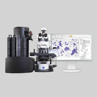
Automatic Cell Imaging System are equipped with multiple modules that coordinate sophisticated tasks such as sample preparation, autofocus mechanisms, fluorescence and brightfield imaging, and multiparametric analysis. These systems excel at multiplexed imaging, where various fluorophores marking different cellular components can be detected simultaneously. This capability is vital for comprehensive cellular characterization since it reveals detailed spatial and temporal interactions within cells and subcellular structures.
Image acquisition hardware generally includes high numerical aperture objectives, sensitive digital cameras, and motorized stage controls that allow systematic scanning of multi-well plates or slides. Alongside advanced optics, integrated software platforms handle image stitching, background correction, feature extraction, and statistical data visualization. User-friendly interfaces cater to both experienced researchers and those new to imaging technology by automating complex imaging protocols, facilitating reproducibility, and minimizing operator errors.
Furthermore, many systems offer live cell incubation chambers maintaining optimal environmental conditions (temperature, CO2, humidity) during prolonged imaging sessions. This feature is indispensable for cell-based assays that require physiological relevance over time. Importantly, the seamless integration of bioinformatics tools enables the linking of imaging data with genomic, proteomic, or metabolomic datasets, fostering multi-omics approaches.
Industrial and Clinical Demands Driving Expansion for Automatic Cell Imaging
The adoption of automatic cell imaging technology is expanding rapidly driven by increasing research funding on personalized medicine, high-throughput screening in pharmaceutical industries, and rising prevalence of chronic diseases requiring cellular-level investigations. The technology has facilitated faster screening of drug candidates by enabling real-time monitoring of drug effects at the single-cell level, thus reducing time-to-market and development costs significantly.
In clinical diagnostics, automatic cell imaging systems are transforming hematology, oncology, and pathology laboratories by improving diagnostic accuracy and enabling quantitative analysis of biopsy samples or blood smears. Early detection of malignancies and infectious diseases is becoming more reliable with precise morphological and phenotypic cellular data. Moreover, the rise of digital pathology combined with image automation is fostering remote diagnostics and telemedicine applications.
The push toward automation in laboratories is also a response to the need for standardized workflows, reduction of human errors, and scaling up of experiments to meet growing data demands. Government initiatives supporting biotechnology innovation and healthcare digitalization further stimulate market penetration.
Navigating Comprehensive Industry Reports on Automatic Cell Imaging Technology Trends
For deeper insights into the evolving landscape of automatic cell imaging systems, detailed market research reports provide invaluable data on emerging trends, technological breakthroughs, vendor analysis, and competitive strategies within the global industry. These reports enable stakeholders to navigate complex market dynamics, appreciate regional and segmental growth opportunities, and gain foresight into investment prospects.
Market intelligence platforms delivering such reports typically feature granular segmentation based on product types, application areas such as drug discovery, cancer research, and regenerative medicine, along with end-user details encompassing research institutions, pharmaceutical companies, and clinical laboratories. These enriched datasets assist companies and researchers in benchmarking technology adoption, understanding regulatory impacts, and identifying partnership opportunities.
Accessing comprehensive industry analyses for automatic cell imaging systems supports informed decision-making, whether for expanding R&D capabilities, entering new markets, or optimizing supply chains. The information therein covers vendor landscapes, revenue forecasts, technology outlook, and innovation pipelines, providing a holistic view of this dynamic sector.
Evolving Innovations Fueling Commercial Viability of Automatic Cell Imaging Devices
Continuous advancements in AI-driven image analysis, integration with lab automation robotics, and development of multiplexed imaging reagents are enhancing the commercial appeal of automatic cell imaging systems. Vendors are increasingly offering customizable platforms tailored to distinct research needs, affordable desktop models for academic labs, and scalable solutions for large pharmaceutical enterprises.
Commercial availability of cloud-based image data management and analytics enhances remote collaboration across global research networks. Moreover, innovations such as label-free imaging and super-resolution microscopy capabilities are addressing limitations in traditional fluorescence-based methods. These improvements broaden application horizons and improve sensitivity and specificity in detecting rare cell populations or subtle phenotypic changes.
The commercial ecosystem also reflects the rising importance of user training, after-sales support, and software updates, as these contribute significantly to customer retention and brand loyalty. Strategic partnerships between imaging technology providers and reagent manufacturers or software developers are another trend shaping the marketplace by offering end-to-end solutions.
Get This Report in Japanese Language: 自動細胞画像化システム
Get This Report in Korean Language: 자동 세포 이미징 시스템
Read More Articles Related to this Industry- Recent developments in Anti Venom Industry
About Author:
Ravina Pandya, Content Writer, has a strong foothold in the market research industry. She specializes in writing well-researched articles from different industries, including food and beverages, information and technology, healthcare, chemical and materials, etc. (https://www.linkedin.com/in/ravina-pandya-1a3984191)