Advanced Applications and Techniques of In-situ Hybridization in Modern Research
In-situ hybridization (ISH) is a powerful molecular biology technique that has revolutionized the way researchers study gene expression, localization of nucleic acids, and chromosomal abnormalities within intact tissues or cells. By utilizing labeled complementary nucleic acid probes to detect specific DNA or RNA sequences, ISH provides spatial context to genetic information, allowing scientists to visualize where genes or transcripts are expressed directly within the histological architecture. This advanced hybridization method has become indispensable for diverse fields such as developmental biology, pathology, cancer research, and clinical diagnostics.
Understanding the Principle and Process of In-situ Hybridization
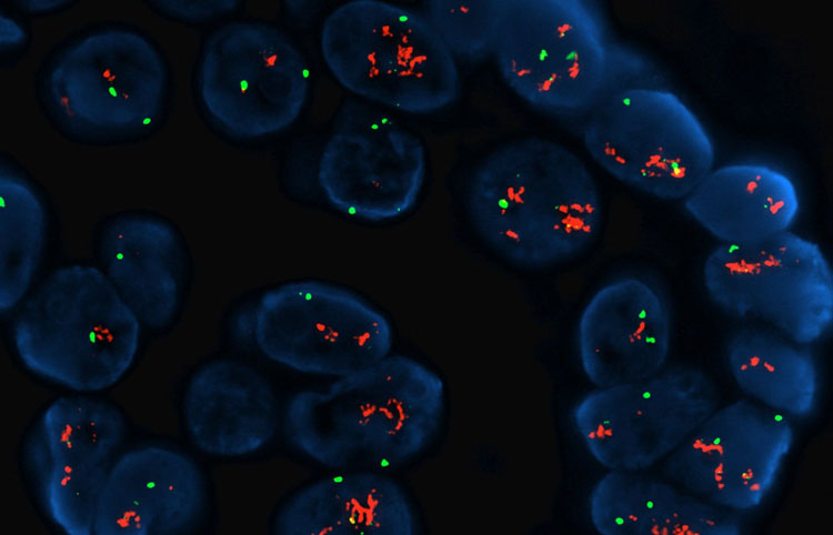
The fundamental principle of In-Situ Hybridization relies on the hybridization of a labeled nucleic acid probe—either DNA or RNA—to its complementary target sequence within fixed cells or tissue sections. The process begins with fixation to preserve tissue morphology and nucleic acid integrity, often using formaldehyde or paraformaldehyde. Subsequently, tissue sections undergo permeabilization to allow probe penetration, followed by hybridization under conditions that favor specific binding between probe and target nucleic acids.
Labeling of probes can be achieved through radioactive isotopes, fluorescent dyes (FISH), or enzyme-linked tags (chromogenic ISH), enabling visualization of hybridized probes via autoradiography, fluorescence microscopy, or bright-field microscopy respectively. After stringent washing to remove nonspecific binding, the hybridized probes are detected, revealing the precise location and abundance of the target sequence within the cellular context.
This technique is highly sensitive and specific, allowing detection of both low-abundance transcripts and chromosomal aberrations. Moreover, the dual capability to probe either RNA or DNA enhances its versatility—RNA ISH informs on gene expression patterns, while DNA ISH assists in identifying structural chromosomal anomalies like translocations, deletions, or amplifications.
In-situ Hybridization Applications Transforming Clinical Diagnostics and Research
The utility of ISH extends widely into clinical diagnostics, where it serves as a pivotal tool for detecting genetic mutations and abnormalities associated with cancer and genetic disorders. Chromogenic ISH and fluorescence ISH (FISH) are standard methods used to identify oncogene amplifications, such as HER2 in breast cancer, or gene fusions like BCR-ABL in chronic myelogenous leukemia.
In infectious disease diagnostics, ISH enables direct localization of pathogen-specific nucleic acids within tissue biopsies, facilitating accurate detection of viral or bacterial infections, even when traditional culture methods fail. This spatial insight differentiates ISH from conventional PCR-based diagnostics by providing valuable histopathological correlates.
In developmental biology and neuroscience, ISH allows researchers to map gene expression patterns during various stages of embryonic development or neural differentiation, offering insights into cellular function and lineage. It is instrumental in investigating how gene malfunction may drive developmental anomalies or neurodegenerative diseases.
Translating ISH into pharmaceutical and biotech research, the method is essential for target validation, enabling the spatial confirmation of gene expression changes in response to drug candidates. This aids in biomarker discovery and therapeutic monitoring, enhancing precision medicine approaches.
Navigating In-situ Hybridization Market Landscape and Industry Trends
The growth of the in-situ hybridization market is driven by the increasing adoption of molecular diagnostic techniques and the rising prevalence of genetic diseases and cancers worldwide. Industry reports analyzing market trends reveal rapid advancements in probe design, automation of ISH procedures, and the integration of multiplexing capabilities to enable simultaneous detection of multiple targets within a single tissue section.
Emerging technologies such as RNAscope and BaseScope have enhanced ISH specificity and sensitivity, enabling researchers and clinicians to detect short RNA targets or splice variants with unprecedented detail. These latest innovations are documented comprehensively in specialized market research reports offering insights into competitive landscapes, technological breakthroughs, and forecasted adoption rates across different regions.
Pharmaceutical companies are also investing heavily in ISH methodologies for companion diagnostic development, which further fuels the demand for refined ISH kits and reagents. Additionally, the implementation of digital pathology and AI-assisted image analysis in ISH data interpretation represents a transformative step toward faster and more accurate diagnosis.
Commercial Outlook of In-situ Hybridization Products and Services
Commercially, the ISH reagents and kits market is expanding alongside growing investments in life science research infrastructure globally. Vendors offering comprehensive ISH solutions—including labeled probes, hybridization buffers, detection systems, and instrumentation—are witnessing heightened demand from academic research institutions, clinical laboratories, and contract research organizations.
The introduction of fully automated ISH platforms is making the technology more accessible to routine clinical laboratories by reducing manual errors, improving throughput, and ensuring reproducibility. This automation, combined with multiplex ISH capability, allows for robust gene expression profiling, which is critical in personalized oncology testing and molecular pathology.
Moreover, the demand for customized probe design services and bioinformatics support is rising, providing end-users with tailored solutions for specific genomic targets. Market insights highlight the growing emphasis on collaboration between reagent manufacturers and diagnostic service providers to enhance ISH test offerings across diverse clinical applications.
Future Advancements Driving In-situ Hybridization Innovation
Looking ahead, innovation in ISH technology will focus on improving multiplexing potential to analyze dozens of RNA targets simultaneously within a single sample, increasing the depth of molecular profiling. Integration of ISH with single-cell sequencing and spatial transcriptomics methods is poised to unlock new dimensions in cellular and tissue-level genetic analysis.
Continuous improvement in probe chemistry, detection sensitivity, and imaging resolution will facilitate the detection of rare transcripts or subtle genetic alterations, essential for early disease diagnosis and monitoring. Furthermore, cross-disciplinary collaborations involving bioinformatics, microscopy, and molecular biology are enabling comprehensive spatially resolved omics approaches.
The in-situ hybridization technique remains at the forefront of molecular diagnostics and research, bridging the gap between genomic data and histopathology. As the demand for personalized medicine and precision diagnostics continues to surge, ISH stands as an invaluable tool transforming biomedical science and healthcare delivery worldwide.
Get this Report in Japanese Language: インサイチューハイブリダイゼーション
Get this Report in Korean Language: 현장 교잡
Read More Articles Related to this Industry
Exploring the Benefits of FTIR Spectroscopy Instruments in Research
About Author:
Money Singh is a seasoned content writer with over four years of experience in the market research sector. Her expertise spans various industries, including food and beverages, biotechnology, chemical and materials, defense and aerospace, consumer goods, etc. (https://www.linkedin.com/in/money-singh-590844163)