Revolutionizing Lung Diagnostics with Endobronchial Ultrasound
Endobronchial ultrasound biopsy (EBUS biopsy) has revolutionized the way pulmonologists diagnose and stage lung diseases, especially lung cancer and mediastinal lymphadenopathy. This minimally invasive procedure utilizes ultrasound technology combined with bronchoscopy to obtain tissue samples from the lungs and surrounding lymph nodes. As advances in medical imaging and diagnostics continue expanding, EBUS biopsy plays a critical role in increasing diagnostic accuracy and improving patient outcomes.
What is Endobronchial Ultrasound Biopsy and How Does it Work?
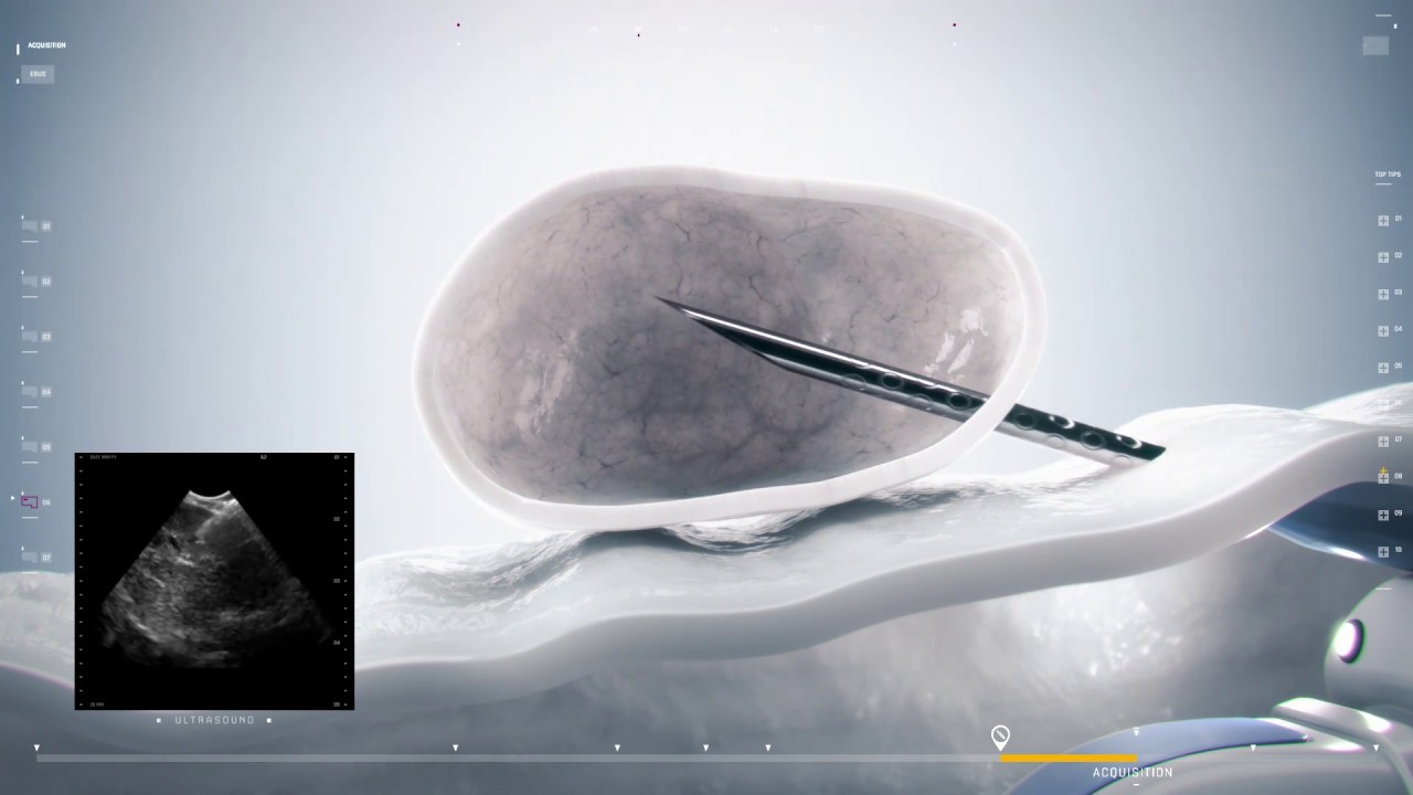
Endobronchial Ultrasound Biopsy is a procedure that uses a specialized bronchoscope equipped with an ultrasound probe at its tip to visualize structures within and adjacent to the airways. The ultrasound probe provides real-time imaging of lymph nodes and masses that lie just outside the airway walls, enabling physicians to guide a needle through the bronchial wall directly into the target lesion for biopsy. This technique allows for the sampling of lymph nodes and masses in the mediastinum and hilum without the need for more invasive surgical methods such as mediastinoscopy or thoracotomy.
The EBUS scope is inserted through the mouth or nose, and once navigated through the tracheobronchial tree, the ultrasound images help identify abnormal tissue. Using transbronchial needle aspiration (TBNA), tissue or fluid samples are collected safely and efficiently. These samples are then sent to pathology for histological and molecular analysis, providing critical information to confirm or rule out cancer, infections, or other pulmonary diseases.
Advantages of Endobronchial Ultrasound Biopsy Over Traditional Techniques
Compared to traditional biopsy methods, EBUS biopsy offers significant advantages regarding invasiveness, diagnostic yield, and patient convenience. Traditional diagnostic modalities like mediastinoscopy require general anesthesia and surgical incisions with longer recovery periods, whereas EBUS biopsy can often be performed under moderate sedation on an outpatient basis.
EBUS biopsy also improves accuracy in staging lung cancer by enabling direct sampling of mediastinal lymph nodes, which is essential before deciding on the appropriate treatment. This minimally invasive approach reduces procedure-related complications, such as bleeding or infection. Furthermore, EBUS biopsy’s real-time imaging capability ensures precise localization, decreasing the chances of inadequate or nondiagnostic samples.
Indications and Clinical Applications of Endobronchial Ultrasound Biopsy
This diagnostic procedure finds wide application in pulmonary medicine, primarily in the evaluation of suspected lung cancer for both diagnosis and staging. It is highly valuable for patients with enlarged mediastinal or hilar lymph nodes detected on computed tomography (CT) or positron emission tomography (PET) scans. Beyond oncology, EBUS biopsy aids in diagnosing infectious diseases like tuberculosis, sarcoidosis, and other granulomatous conditions by providing tissue samples for microorganism identification and histopathological analysis.
In addition, EBUS biopsy is used to investigate unexplained mediastinal lymphadenopathy, lymphoma, and metastatic diseases originating outside the lungs. This versatility establishes EBUS as an essential tool in the diagnostic armamentarium, providing critical information that guides personalized treatment plans and reduces the need for more invasive surgeries.
How to Access the Latest Market Insights on Endobronchial Ultrasound Biopsy
For healthcare providers, medical device companies, and investors seeking in-depth data on the endobronchial ultrasound biopsy market landscape, recent research reports highlight technological trends, growth drivers, competitive dynamics, and future opportunities. These comprehensive market research reports analyze key factors such as product innovations, emerging applications, regulatory frameworks, and regional market performance.
Detailed segmentation covers product types, modality expansions, and end-user profiles including hospitals, outpatient clinics, and specialty centers. The reports also forecast market trajectories based on historical data and predictive analytics, offering valuable insights for strategic decision-making and business development. Accessing this intelligence supports better understanding of market evolution, investment prospects, and commercialization strategies.
Equipment and Consumables Utilized in Endobronchial Ultrasound Biopsy Procedures
Executing EBUS biopsy requires specialized equipment designed to combine bronchoscopy with high-frequency ultrasound imaging. The core device, an EBUS bronchoscope, is a flexible endoscope integrated with a convex ultrasound transducer at its distal tip. This allows visualization beyond the airway walls while maintaining maneuverability through bronchial passages.
In conjunction with the bronchoscope, dedicated needles and aspiration kits are essential for obtaining cellular samples. Needles vary in gauge and length, tailored for different tissue targets. Accessories such as biopsy brushes, cytology brushes, and sample preservation solutions complement the procedure by ensuring specimen integrity.
Additionally, integrated ultrasound processors and video processors provide clear imaging and recording capabilities, assisting pulmonologists in performing accurate and efficient biopsies. The synergy of advanced equipment and consumables underpins the success and safety of EBUS biopsy interventions.
Future Trends Driving Growth in Endobronchial Ultrasound Biopsy Market
The endobronchial ultrasound biopsy market is anticipated to grow steadily due to increasing prevalence of lung cancer and respiratory disorders worldwide. Continuous technological advancements, including the integration of robotic assistance and enhanced imaging modalities, are expected to expand procedural capabilities and accuracy.
Emerging trends focus on miniaturization of devices, improved needle design for better tissue yield, and incorporation of artificial intelligence to aid image interpretation. Increasing awareness about minimally invasive diagnostic alternatives and favorable reimbursement policies are also promoting wider adoption.
Furthermore, growing demand in emerging economies, coupled with expanding geriatric populations prone to chronic respiratory diseases, is stimulating market growth. Collaborations between medical device manufacturers and healthcare providers are fostering innovation, thereby opening new avenues for clinical application and commercial success.
Get this Report in Japanese Language: 気管支内超音波生検市場
Get this Report in Korean Language: 기관지 내 초음파 생검 시장
About Author:
Money Singh is a seasoned content writer with over four years of experience in the market research sector. Her expertise spans various industries, including food and beverages, biotechnology, chemical and materials, defense and aerospace, consumer goods, etc. (https://www.linkedin.com/in/money-singh-590844163)